Early-systolic shape
Upstroke, slope, and template similarity propose an opening-related landmark.
Radar Signal Processing · Multimodal Cardiac Research
A research prototype that aligns ECG electrical anchors, SCG mechanical references, and non-contact FMCW radar morphology on a beat-relative axis for AO/AC candidate and PEP/LVET/QS2 analysis.
Actual Experiment Evidence
These user-captured photographs show the actual ECG, SCG, and FMCW radar hardware and measurement setup. They are not AI-generated or composited equipment; public derivatives use only orientation correction, deterministic cropping, resizing, and privacy blur.
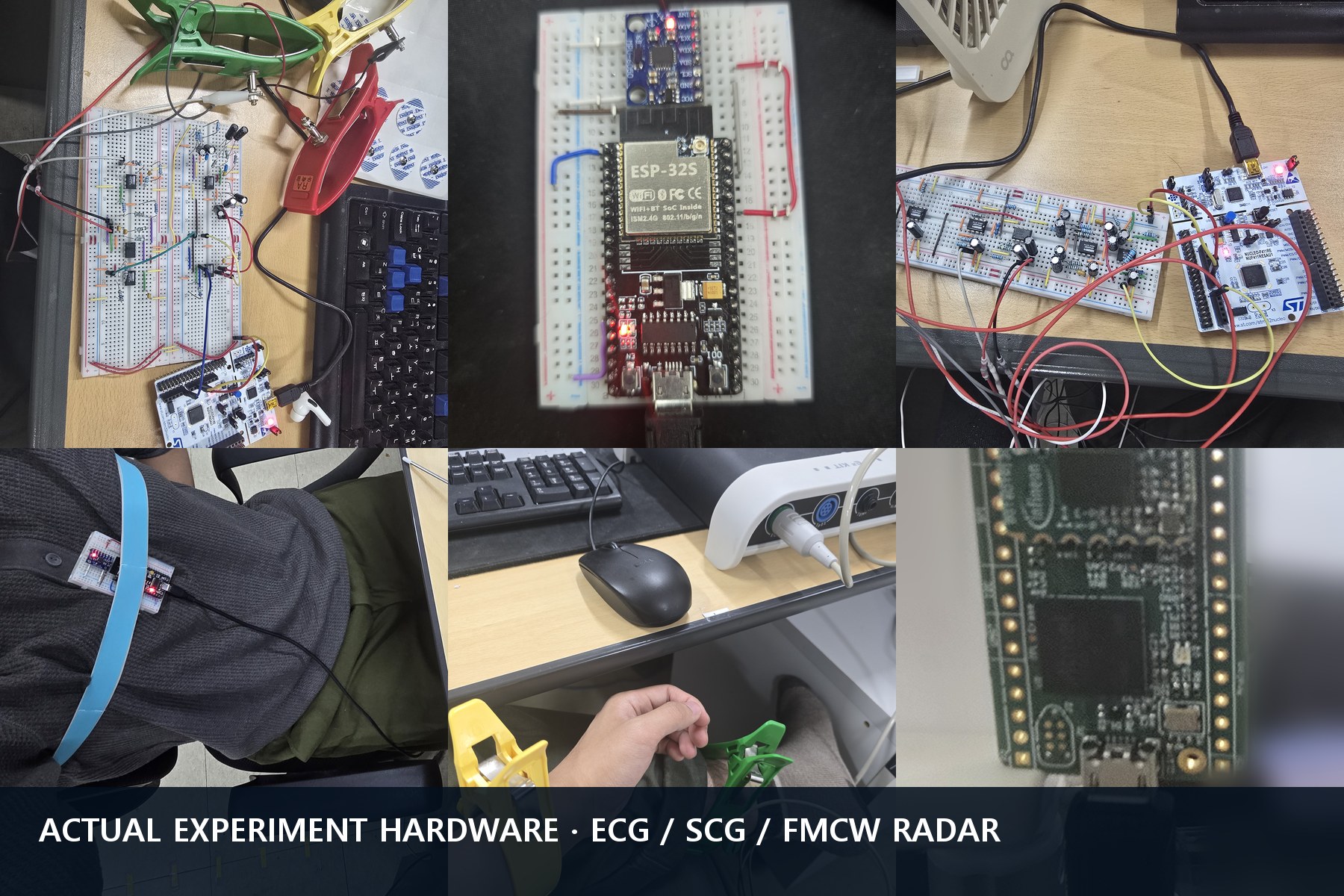
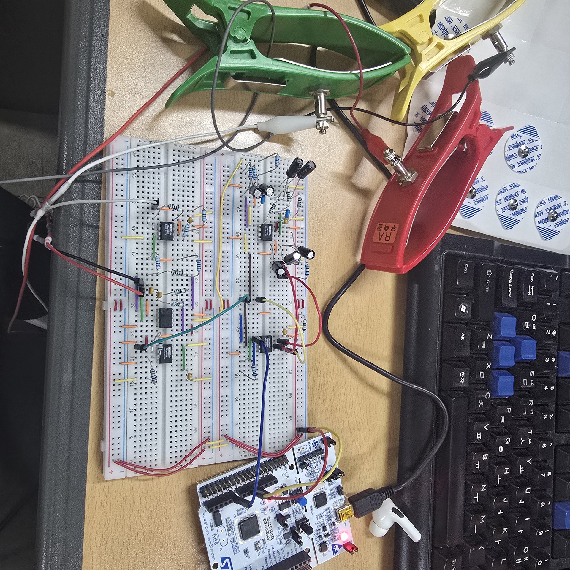
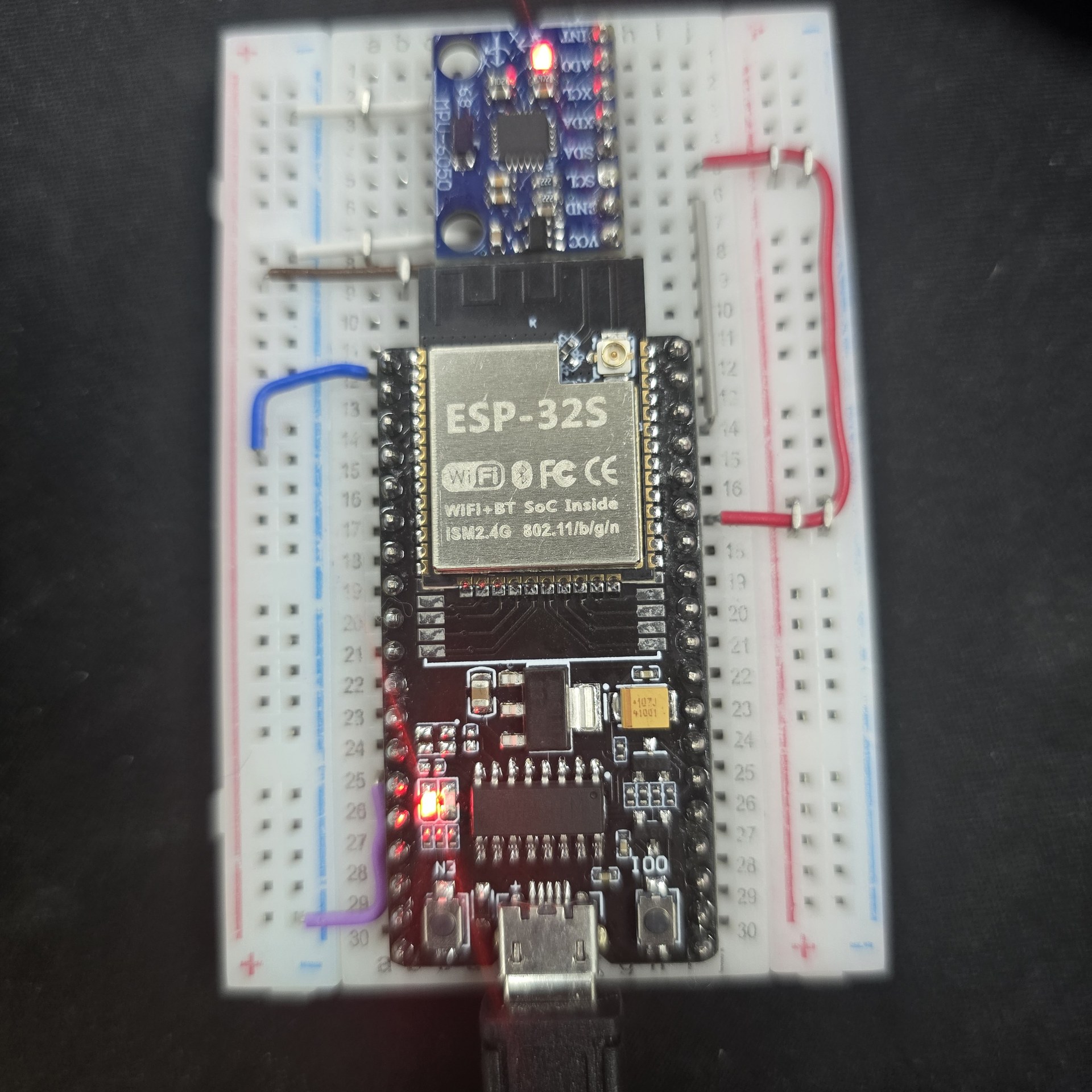
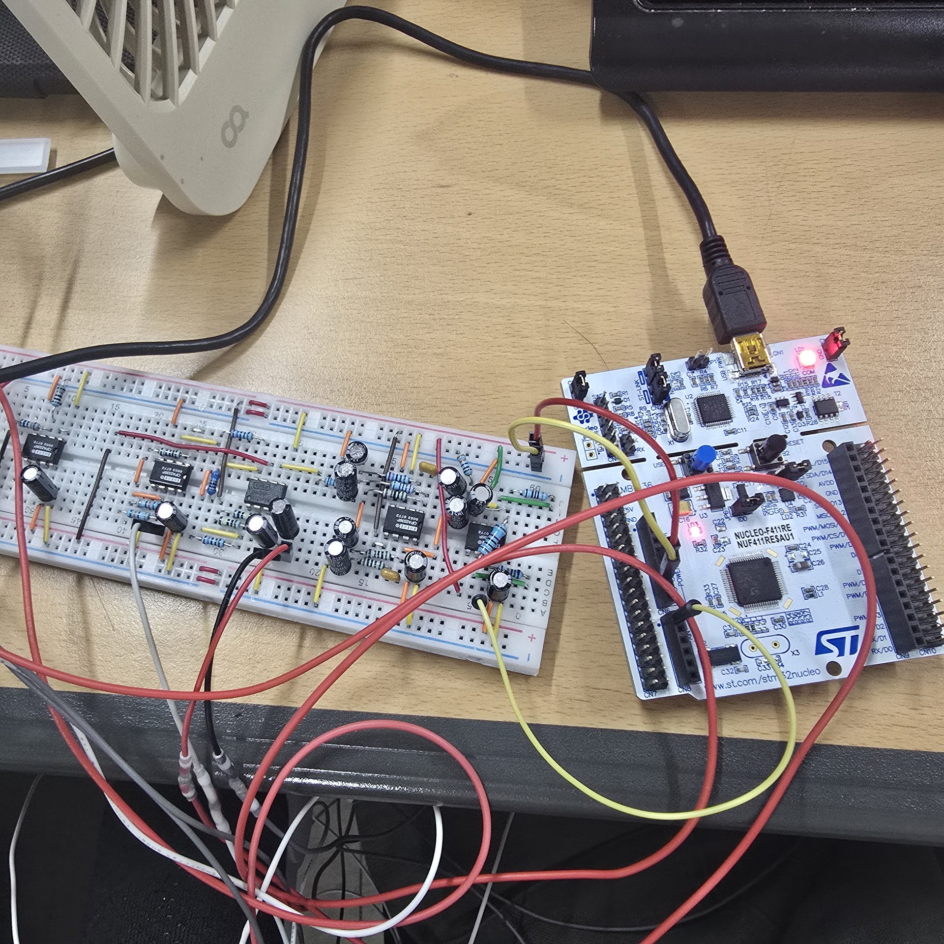
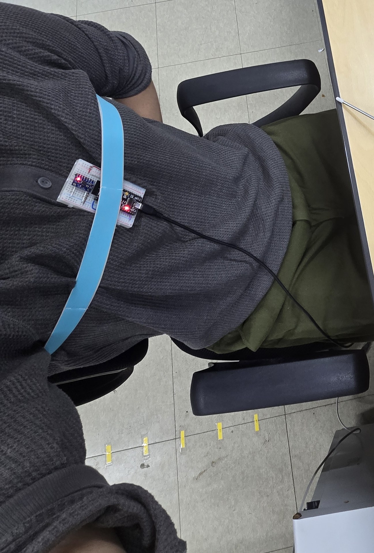
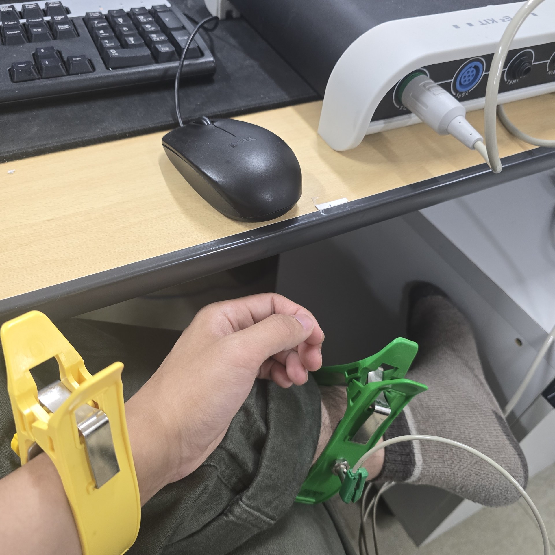
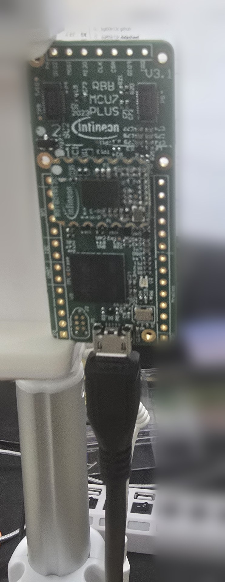
01 · Radar Path
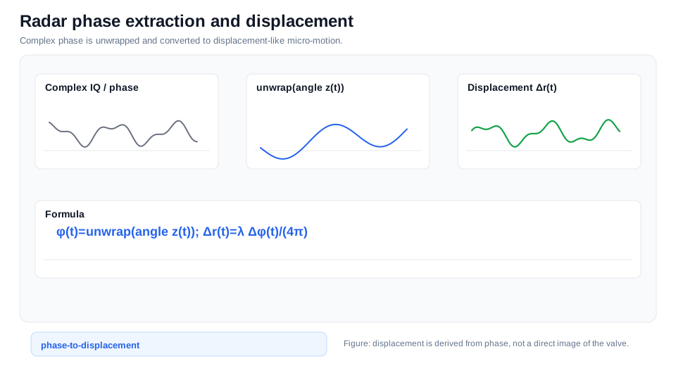
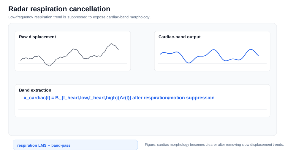
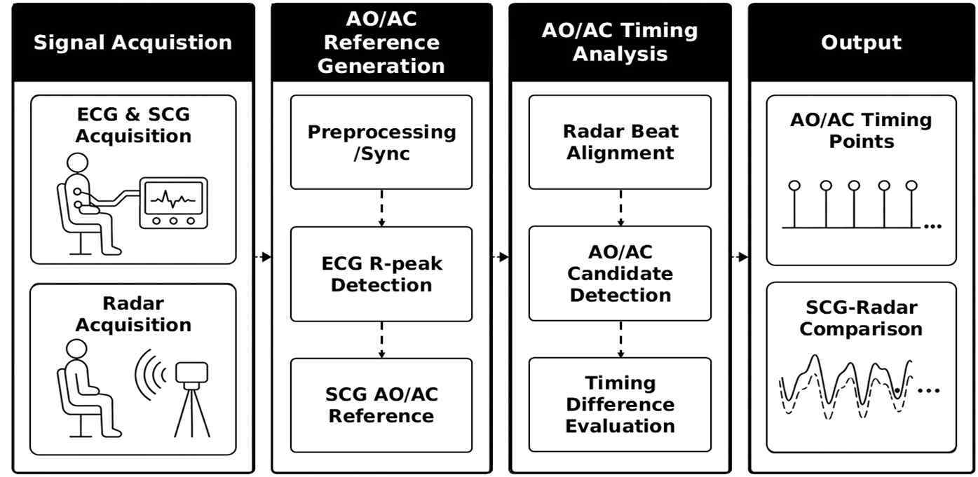
The pipeline selects a region of interest after range FFT, extracts phase displacement, and suppresses respiration/motion components before beat analysis.
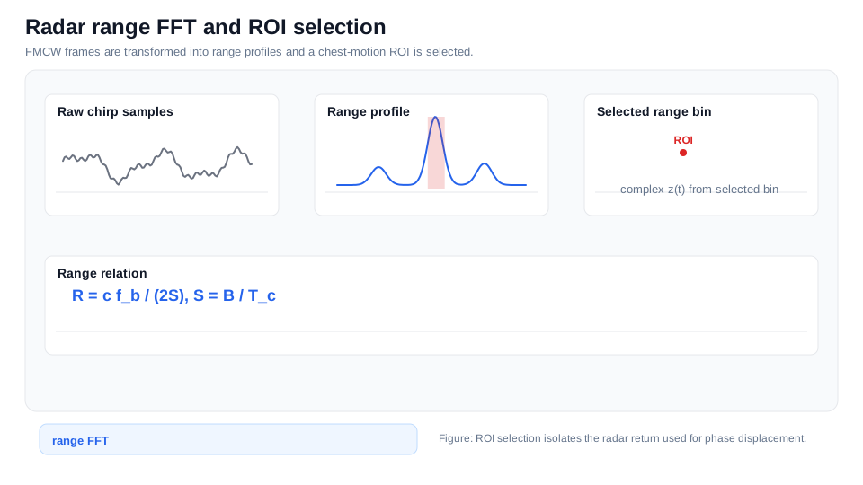
02 · Alignment
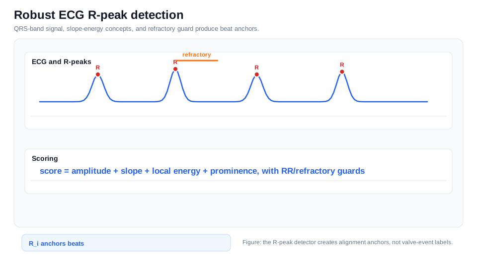
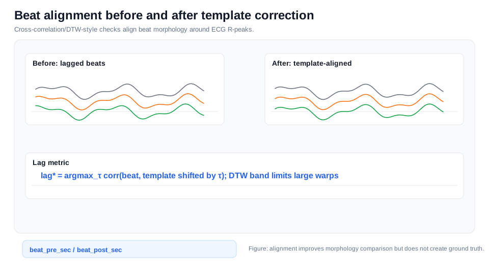
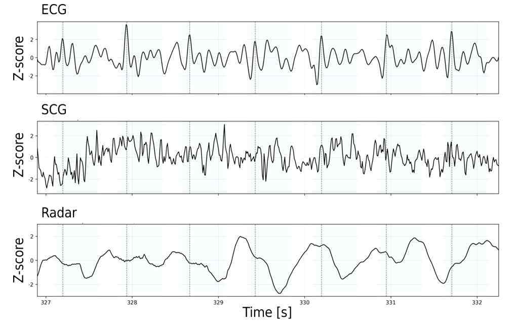
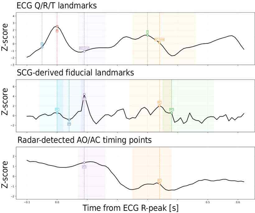
ECG R-peaks provide stable beat anchors; SCG references and radar morphology are compared on the same relative time axis.
03 · Detection
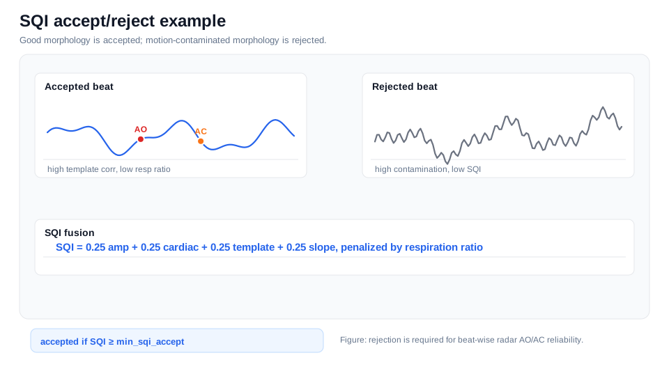
Slope, morphology, template, wavelet, and notch detectors contribute timing candidates; confidence and SQI reject unstable beats.
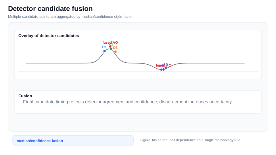
Upstroke, slope, and template similarity propose an opening-related landmark.
Notch, inflection, and temporal continuity track a closure-related candidate.
Beat quality and detector agreement make both outputs and exclusion reasons inspectable.
04 · Paper-source evidence
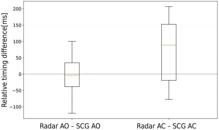
The paper PDF was audited at the embedded-image level. Four substantive figures are public; three companion mask layers were not duplicated. No participant-level raw data or subject CSV is included.
05 · Evidence Boundary
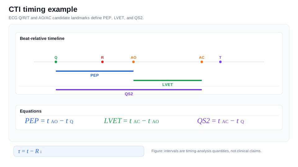
The repository includes analysis code, firmware, configurations, synthetic algorithm diagrams, and four paper-source figures. It does not publish participant raw data or subject-level result CSV, and it makes no clinical performance claim.
| Item | Repository state | Meaning |
|---|---|---|
| Analysis pipeline | Implemented | Python processing, detectors, SQI, and export documented |
| Embedded acquisition | Documented | STM32 ECG and ESP32 MPU6050 SCG firmware included |
| Public raw data | Not included | Only synthetic explanatory figures are public |
| Absolute valve-event validation | Future validation | Requires independent echo, ICG, or PCG reference |